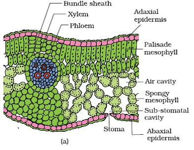
The Lamina part of the leaf when used for taking longitudinal section will show three major divisions called as epidermal layer, mesophyll layer and vascular system. Epidermis layer will be present on the upper as well as lower surfaces of the leaf while these layers are covered by another thin casing called as cuticle. The stomata number are found to be more in the lower epidermis than in the upper. Sometimes, the upper epidermal layer might not have stomata at all. The area that is present between the upper and lower epidermis is filled by a tissue known as mesophyll. The mesophyll tissue consists of chloroplasts which help the cells to carryout the process of photosynthesis. The mesophyll tissue comprises of parenchyma cells. This parenchyma of mesophyll is divided into two types. They are palisade and spongy parenchyma. The palisade layer that is present towards the upper epidermis comprises of cells that are elongated. The palisade cells are arranged parallel to one another and are arranged vertically. The oval shaped or round shaped tissue that is present below the palisade parenchyma layer is spongy parenchyma. This tissue spreads towards the lower epidermal layer. The spongy parenchyma is intervened by the air cavities that are bigger in size. The vascular system is made up of vascular bundles which are visible in the midrib region as well as in the veins. The vein size is dependent on the vascular bundle size. The thickness of the veins in the leaf differs at various regions in a dicot leaf. There is a layer of cells present covering the vascular bundles. This layer is made up of bundle sheath cells.
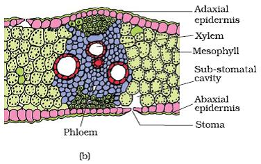
Isobilateral Leaf-Monocot Leaf
In an isobilateral leaf, unlike in dorsiventral leaf the mesophyll is not differentiated into palisade and spongy parenchyma. The stomata in isobilateral leaf are found to be present on both the surfaces of the leaf. In the case of the plants that belong to the grass family, some of the upper epidermal cells present all along the veins are found to be bigger, colorless and empty. These cells are termed as bulliform cells. If the bulliform cells are turgid and filled with water, the surface of the leaf will remian exposed. If the leaves are flaccid due to the water stress, the leaves become curled towards inside to reduce the water loss. The small size of the vascular bundles appearing in the cross section of monocot leaves indicate the presence of parallel venation.
T.S. of dorsiventral Dicot Leaf
No comments:
Post a Comment